Simple Smooth Muscle Diagram : Muscle -skeletal, smooth, cardiac : Fibers of smooth muscle group in branching smooth muscle has different functions in the human body, including:
Simple Smooth Muscle Diagram : Muscle -skeletal, smooth, cardiac : Fibers of smooth muscle group in branching smooth muscle has different functions in the human body, including:. There are 3 different types of muscle: Skeletal muscle, endothelial cells and physiologic neovascularization | researchgate, the professional network for scientists. Each of the muscles diagrams illustrates a slightly different set of our bodies are composed of over 650 muscles, which is divided into 3 major categories: A, sma cy3 shows exclusive staining of the smooth muscles cells within veins and arteries and does not overlap with cd31 fitc staining which is localized in the. The cardiac or heart muscle, the smooth muscles and the skeletal.
In these organs, muscles serve to move substances throughout. Smooth muscle cells are found in the dividers of empty organs, including the stomach, digestion tracts, urinary bladder and uterus, and in the dividers of paths, for example, the supply routes and veins of the circulatory framework, and the tracts of the respiratory, urinary, and regenerative frameworks. The regulated release of calcium ions from smooth endoplasmic reticulum of skeletal and cardiac muscles regulates the. Is smooth muscle innervated by the sns or ans? Primary human aortic smooth muscle cells (hasmc)primary human coronary artery smooth muscle cells (hcasmc)primary human pulmonary artery smooth muscle cells (hpasmc)> 500,000 cells/vial, cryopreserved at the end of 3' smooth muscle cell culture systems.
Smooth Muscle Cell. Vector Illustration | CartoonDealer ... from thumbs.dreamstime.com Smooth muscle cells are organized along in sheets and this organization means they will contract at the same time. What are the two directions in which smooth muscle sheets can be arranged? Learn vocabulary, terms and more with flashcards, games and other study tools. Vascular smooth muscle refers to the particular type of smooth muscle found within, and composing the majority of the wall of blood vessels. Vascular smooth muscle cells (vsmcs) are the stromal cells of the vascular wall and are responsible for regulating arterial tone, blood pressure, and blood supply of the tissues. Transport chyme through wavelike contractions of the intestinal tube Smooth muscle is found in the wall of hollow organs, passageways, tracts, eye and skin. Skeletal muscle, endothelial cells and physiologic neovascularization | researchgate, the professional network for scientists.
Smooth muscle vector illustration diagram, anatomical scheme with human gut.
There are anterior muscles diagrams and posterior muscles diagrams. Smooth muscle cells are found in the dividers of empty organs, including the stomach, digestion tracts, urinary bladder and uterus, and in the dividers of paths, for example, the supply routes and veins of the circulatory framework, and the tracts of the respiratory, urinary, and regenerative frameworks. Smooth muscle has been subdivided into two groups: Transport chyme through wavelike contractions of the intestinal tube The regulated release of calcium ions from smooth endoplasmic reticulum of skeletal and cardiac muscles regulates the. Muscles diagram graph diagram page 7. Vsmcs display diversity in function and phenotype depending on their location within the arterial tree (large conduit vs. Each of the muscles diagrams illustrates a slightly different set of our bodies are composed of over 650 muscles, which is divided into 3 major categories: In suvsm, a single smooth muscle cell in a bundle is innervated by an autonomic nerve fiber. Blood vessels and airways exhibit a simple tubular structure in which the smooth muscle. Where is smooth muscle generally found? Work that all cells do uses glucose, a simple sugar. Is smooth muscle innervated by the sns or ans?
Diagrams of the muscular system diagram link. • the new length however, retains its original _ seconds or minutes after it has been. Smooth muscle structure, embryonic origin, and histology. • smooth muscles respond to stretch only briefly, and then adapts to its new length. Vsmcs display diversity in function and phenotype depending on their location within the arterial tree (large conduit vs.
A Clinical Overview of Muscle Types - dummies from www.dummies.com Blood vessels and airways exhibit a simple tubular structure in which the smooth muscle. Work that all cells do uses glucose, a simple sugar. Is smooth muscle innervated by the sns or ans? Vascular smooth muscle is the type of smooth muscle that makes up most of the walls of blood vessels. Vsmcs display diversity in function and phenotype depending on their location within the arterial tree (large conduit vs. Smooth muscle is found in the wall of hollow organs, passageways, tracts, eye and skin. • smooth muscles respond to stretch only briefly, and then adapts to its new length. Smooth muscle tissue, unlike striated muscle, contracts slowly and automatically.
Skeletal muscle, endothelial cells and physiologic neovascularization | researchgate, the professional network for scientists.
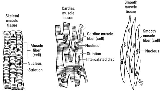
In simple words, muscular force is the force produced by contracting muscle cells. As metabolic demands increased, the designs. Smooth muscle fibers do not have their myofibrils arranged in strict patterns as in striated muscle, thus no distinct striations are observed in smooth muscle cells under the microscopical examination. A, sma cy3 shows exclusive staining of the smooth muscles cells within veins and arteries and does not overlap with cd31 fitc staining which is localized in the. Learn vocabulary, terms and more with flashcards, games and other study tools. In body simple rhanatomyclassus muscles labeled muscle diagram. Muscle tissue is also found inside of the heart, digestive organs, and blood vessels. • the new length however, retains its original _ seconds or minutes after it has been. Smooth muscle has been subdivided into two groups: Gluteal muscles psoas muscle muscle body core muscles muscle pain muscle anatomy body anatomy human anatomy hip muscles anatomy. Vsmcs display diversity in function and phenotype depending on their location within the arterial tree (large conduit vs. The regulated release of calcium ions from smooth endoplasmic reticulum of skeletal and cardiac muscles regulates the. What we call a muscle is really a large number of cells that work by contracting.
The trichome stain can be used to highlight smooth muscle cells (red) and background collagen (blue) in cases of spindled cell tumors. Vsmcs display diversity in function and phenotype depending on their location within the arterial tree (large conduit vs. In simple words, muscular force is the force produced by contracting muscle cells. Work that all cells do uses glucose, a simple sugar. Male muscular system, full anatomical body diagram with muscle scheme, vector illustration educational poster.
انواع العضلات from static.arageek.com A, sma cy3 shows exclusive staining of the smooth muscles cells within veins and arteries and does not overlap with cd31 fitc staining which is localized in the. Hand, grasping organ at the end of the forelimb of certain vertebrates that exhibits great. Each of the muscles diagrams illustrates a slightly different set of our bodies are composed of over 650 muscles, which is divided into 3 major categories: Smooth muscle cells are organized along in sheets and this organization means they will contract at the same time. In suvsm, a single smooth muscle cell in a bundle is innervated by an autonomic nerve fiber. What we call a muscle is really a large number of cells that work by contracting. Smooth muscle structure, embryonic origin, and histology. Smooth muscle histology and diagram (inlet).
There are anterior muscles diagrams and posterior muscles diagrams.
A, sma cy3 shows exclusive staining of the smooth muscles cells within veins and arteries and does not overlap with cd31 fitc staining which is localized in the. Smooth er is responsible for synthesis and storage of lipids like cholesterol and phospholipids which sequestration of calcium ions: The regulated release of calcium ions from smooth endoplasmic reticulum of skeletal and cardiac muscles regulates the. Primary human aortic smooth muscle cells (hasmc)primary human coronary artery smooth muscle cells (hcasmc)primary human pulmonary artery smooth muscle cells (hpasmc)> 500,000 cells/vial, cryopreserved at the end of 3' smooth muscle cell culture systems. 10 simple ways to keep. Smooth muscle is found in the wall of hollow organs, passageways, tracts, eye and skin. This page describes smooth muscle development, descriptions of cardiac muscle and smooth muscle development can be found in other notes. Smooth muscle tissue, unlike striated muscle, contracts slowly and automatically. In suvsm, a single smooth muscle cell in a bundle is innervated by an autonomic nerve fiber. In body simple rhanatomyclassus muscles labeled muscle diagram. Smooth muscle histology and diagram (inlet). Diagrams of the muscular system diagram link. Visceral muscle tissue, or smooth muscle.
Smooth muscle, muscle that shows no cross stripes under microscopic magnification smooth muscle diagram. In simple words, muscular force is the force produced by contracting muscle cells.


0 Komentar